Heading
For Doctor's Appointment,
Click Here for Online Appointments (or)
Call us @ + 91 98414 97037
For Doctor's Appointment,
Click Here for Online Appointments (or)
Call us @ + 91 98414 97037

Solving all kind of visual problems.

Solving all kind of visual problems.

Solving all kind of visual problems.

Solving all kind of visual problems.

Retinal Service provides highly specialized diagnosis and treatment of complex diseases of the retina...
more.png)
LASIK is a surgical procedure that uses laser to correct nearsightedness, farsightedness, and/or astigmatism.
more
Opacification of transparent lens in our eye is called Cataract. The cataractous lens is broken ...
moreParagraph

 Caring for your vision doesn't begin and end with eyeglasses, contact lenses and corneal modification surgeries like LASIK. There are many other things you can do to maximize and protect the vision you
currently have. An essential component of healthy vision is a yearly comprehensive eye examination. Optometrists detect and treat problems and conditions of the eye and can help you get the most from your vision. Optometrists can
also diagnose systemic health problems that require treatment by your primary care physician. Periodic eye and vision examinations are an important part of preventive health care. Many eye and vision problems have no obvious signs
or symptoms, so you might not know a problem exists.
Caring for your vision doesn't begin and end with eyeglasses, contact lenses and corneal modification surgeries like LASIK. There are many other things you can do to maximize and protect the vision you
currently have. An essential component of healthy vision is a yearly comprehensive eye examination. Optometrists detect and treat problems and conditions of the eye and can help you get the most from your vision. Optometrists can
also diagnose systemic health problems that require treatment by your primary care physician. Periodic eye and vision examinations are an important part of preventive health care. Many eye and vision problems have no obvious signs
or symptoms, so you might not know a problem exists.
Early diagnosis and treatment of eye and vision problems can help prevent vision loss. Each patient's signs and symptoms, along with your optometrist's professional judgment, will determine what tests your optometrist conducts.
| Age | Risk Free | At Risk |
|---|---|---|
| Birth to 24 Months | At 6 months of age | By 6 months of age or as recommended |
| 2 to 5 years | At 3 years of age | At 3 years of age or as recommended |
| 6 to 60 years | Every two years | Every one to two years or as recommended |
| 61 and older | Annually | Annually or as recommended |
 Kids are always special to us. So is theirvision more precious. If a kid cannot see clearly, they do not realize it andthey think everybody sees like that. If you are a spec wearer
or ask yourfriend who has specs, they will acknowledge the same.
Kids are always special to us. So is theirvision more precious. If a kid cannot see clearly, they do not realize it andthey think everybody sees like that. If you are a spec wearer
or ask yourfriend who has specs, they will acknowledge the same.
All kids need to be checked around 5 yearseven if they do not have visual complaint. Ideally In a clinic with individualattention. Around 30-40 % of kids need specs. Around 10% kids never realizethat they need to have specs. 1-2% become amblyopic (lazy eye).
Squint is another common pediatric problemwe have a solution for it. We have Orthoptics Exercises to Surgical interventionas treatment options.
 A cataract is a clouding of the lens in the eye which leads to a decrease in vision. Cataracts often develop slowly and can affect one or both eyes. Symptoms may include faded colors,
blurry vision, halos around light, trouble with bright lights, and trouble seeing at night.
A cataract is a clouding of the lens in the eye which leads to a decrease in vision. Cataracts often develop slowly and can affect one or both eyes. Symptoms may include faded colors,
blurry vision, halos around light, trouble with bright lights, and trouble seeing at night.
Opacification of the transparent lens in our eye is called CATARACT. The Cataractous lens is broken into multiple pieces, emulsified and sucked out of the eye through a micro incision followed with an Artificial Intra Ocular Lens (IOL) is Implantation.
State of art cataract surgery is available with us. Microincision Phacoemulsification cataract surgery with foldable IOLS to LASER CATARACT SURGREY for a speedy recovery.

A variety of options are available for IntraOcular Lenses. A detailed examination and discussion will allow for the bestpossible option for every individual patient. Customized services in helping the patientto restore and add value to vision with ultra modern technology has always keptour patients happy.

 Glaucoma is known as a silent killer of the eye as the patient loses vision over a period of time without their knowledge. In this condition, the Intraocular Pressure is more for the
Eye, which gradually compromises the nutrient supply to theOPTIC NERVE (the nerve responsible for vision) which eventually leads to decrease in vision. As the nerve is affected, It can lead to INCURABLE BLINDNESS.
Glaucoma is known as a silent killer of the eye as the patient loses vision over a period of time without their knowledge. In this condition, the Intraocular Pressure is more for the
Eye, which gradually compromises the nutrient supply to theOPTIC NERVE (the nerve responsible for vision) which eventually leads to decrease in vision. As the nerve is affected, It can lead to INCURABLE BLINDNESS.
So the key lies in EARLY DETECTION. Glaucoma is detected through a comprehensive dilated eye exam that includes the following:
A spectrum of treatment modalities areavailable for Glaucoma. Your doctor will decide on the best depending on your condition.
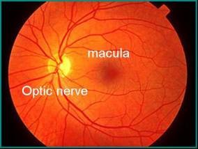
Chronically high blood sugar from diabetes is associated with damage tothe tiny blood vessels in the retina, leading to diabetic retinopathy. Theretina detects light and converts it to signals sent through the optic nerve tothe brain. Diabetic retinopathy can cause blood vessels in the retina to leakfluid or hemorrhage (bleed), distorting vision. In its most advanced stage, newabnormal blood vessels proliferate (increase in number) on the surface of theretina, which can lead to scarring and cell loss in the retina.

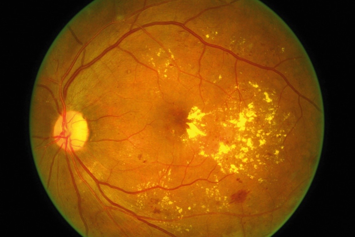

 PDR is the more advanced form of the disease. At this stage, circulation problems deprive the retina of oxygen. As a result new, fragile blood vessels can begin to grow in the retina and into the vitreous,
the gel-like fluid that fills the back of the eye. The new blood vessels may leak blood into the vitreous, clouding vision.
PDR is the more advanced form of the disease. At this stage, circulation problems deprive the retina of oxygen. As a result new, fragile blood vessels can begin to grow in the retina and into the vitreous,
the gel-like fluid that fills the back of the eye. The new blood vessels may leak blood into the vitreous, clouding vision.
Diabetic Macular Edemais the build-up of fluid (edema) in a region of the retina called the macula.The macula is important for the sharp, straight-ahead vision that is used forreading, recognizing faces, and driving. DME is the most common cause of visionloss among people with diabetic retinopathy.
About half of all people withdiabetic retinopathy will develop DME. Although it is more likely to occur asdiabetic retinopathy worsens, DME can happen at any stage of the disease.
People with all typesof diabetes (type 1, type 2, and gestational) are at risk for diabetic retinopathy. Riskincreases the longer a person has diabetes. Between 40 and 45 percent of peoplediagnosed with diabetes have some stage of diabetic retinopathy, although onlyabout half are aware of it. Women who develop or have diabetes during pregnancymay have rapid onset or worsening of diabetic retinopathy.
Longer the DiabeticLife Higher the risk. When the Sugar controlis stable, continuous then the risk is less.


The same scene as viewed by a person normal vision (Left) and with(Center) advanced diabetic retinopathy. The floating spots are hemorrhages thatrequire prompt treatment. DME (Right) causes blurred vision.
The early stages ofdiabetic retinopathy usually have no symptoms. The disease often progressesunnoticed until it affects vision. Bleeding from abnormal retinal blood vesselscan cause the appearance of “floating” spots. These spots sometimes clear ontheir own. But without prompt treatment, bleeding often recurs, increasing therisk of permanent vision loss. If DME occurs, it can cause blurred vision.
No.21, Nathan's Arcade,
Opp. HDFC Bank, L.B. Road,
Thiruvanmiyur, Chennai,
Tamil Nadu 600041

To Top